
Intro
Discover the ins and outs of a Transesophageal Echocardiogram (TEE) - a specialized ultrasound test that provides clear images of the heart and its blood vessels. Learn about the procedure, preparation, risks, and benefits of TEE, as well as its uses in diagnosing heart conditions such as atrial fibrillation, heart valve disease, and cardiac tumors.
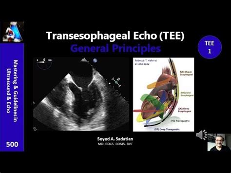
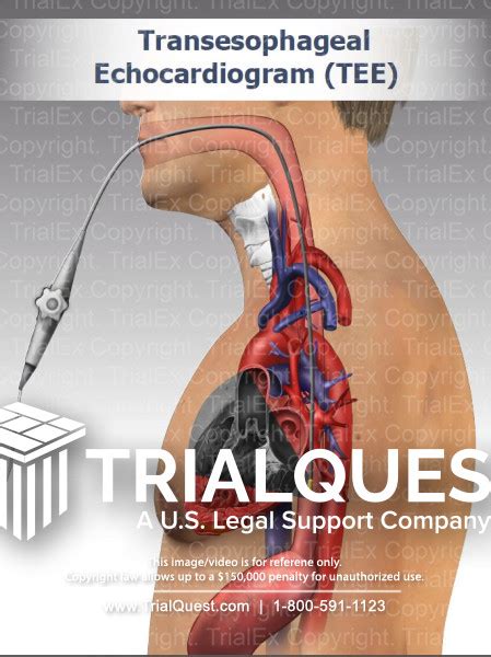
A transesophageal echocardiogram, commonly referred to as a TEE, is a specialized medical imaging test that uses high-frequency sound waves to produce detailed images of the heart and its surrounding structures. Unlike a traditional echocardiogram, which is performed through the chest wall, a TEE involves inserting an ultrasound probe through the mouth and into the esophagus, providing a closer and more detailed view of the heart.

The Importance of TEE in Cardiac Care
A TEE is a valuable diagnostic tool that offers several advantages over traditional echocardiography. Its ability to provide high-resolution images of the heart's structures and functions makes it an essential test for diagnosing and managing various cardiac conditions. Some of the key benefits of TEE include:
- Improved image quality: The proximity of the ultrasound probe to the heart allows for more detailed and accurate images, which can help doctors diagnose conditions that may not be visible through traditional echocardiography.
- Enhanced diagnostic accuracy: TEE can help doctors diagnose conditions such as heart valve problems, cardiac tumors, and blood clots, which can be difficult to detect through other imaging tests.
- Guiding interventional procedures: TEE can be used to guide doctors during interventional procedures, such as heart valve repairs or replacements, by providing real-time images of the heart.
How TEE is Performed
A TEE is typically performed in a hospital or outpatient setting by a trained healthcare professional. The test involves the following steps:
- Preparation: The patient is asked to fast for several hours before the test and may be given medication to help them relax.
- Insertion of the ultrasound probe: The patient is asked to lie on their side, and the ultrasound probe is inserted through their mouth and into their esophagus.
- Imaging: The ultrasound probe sends high-frequency sound waves through the esophagus and into the heart, producing detailed images of the heart's structures and functions.
- Data analysis: The images are analyzed by a cardiologist or other trained healthcare professional to diagnose and manage cardiac conditions.
Benefits of TEE
TEE offers several benefits over traditional echocardiography, including:
Improved diagnostic accuracy
TEE can help doctors diagnose conditions that may not be visible through traditional echocardiography.
Guiding interventional procedures
TEE can be used to guide doctors during interventional procedures, such as heart valve repairs or replacements.
Monitoring cardiac function
TEE can be used to monitor cardiac function during surgery or other medical procedures.
Risks and Complications of TEE
While TEE is generally a safe test, there are some potential risks and complications to be aware of, including:
- Discomfort: Some patients may experience discomfort or difficulty swallowing during the test.
- Bleeding: There is a small risk of bleeding or injury to the esophagus or surrounding tissues.
- Infection: There is a small risk of infection with any invasive medical procedure.

Preparing for a TEE
To prepare for a TEE, patients should:
- Fast: Patients should fast for several hours before the test.
- Avoid medications: Patients should avoid taking certain medications, such as blood thinners, before the test.
- Inform their doctor: Patients should inform their doctor of any medical conditions or concerns they may have.
Conclusion
A transesophageal echocardiogram is a valuable diagnostic tool that offers several advantages over traditional echocardiography. Its ability to provide high-resolution images of the heart's structures and functions makes it an essential test for diagnosing and managing various cardiac conditions. While there are some potential risks and complications to be aware of, the benefits of TEE make it a valuable tool in cardiac care.
FAQs
What is a transesophageal echocardiogram?
+A transesophageal echocardiogram is a medical imaging test that uses high-frequency sound waves to produce detailed images of the heart and its surrounding structures.
What are the benefits of TEE?
+TEE offers several benefits, including improved diagnostic accuracy, guiding interventional procedures, and monitoring cardiac function.
What are the risks and complications of TEE?
+While TEE is generally a safe test, there are some potential risks and complications to be aware of, including discomfort, bleeding, and infection.